Retina consults in under 60 seconds.
Capture OCT, OCT-A and fundus images straight from your browser, ask one clear question, and get a signed interpretation from a credentialed retina specialist. Built for optometrists and emergency departments.
0:03
Source selected
Your OCT capture source is remembered from last time.
0:31
Question asked
OD, new distortion for 3 days. Fluid? How soon to refer?
0:12
Six frames captured
Press Space per frame. Thumbnails upload while you keep going.
0:44
Consult submitted
One credit reserved. Eligible retina specialists see it now.
0:03
Source selected
Your OCT capture source is remembered from last time.
0:12
Six frames captured
Press Space per frame. Thumbnails upload while you keep going.
0:31
Question asked
OD, new distortion for 3 days. Fluid? How soon to refer?
0:44
Consult submitted
One credit reserved. Eligible retina specialists see it now.
What you send, and what comes back
Synthetic example. Not a real patient.
You send
You get back
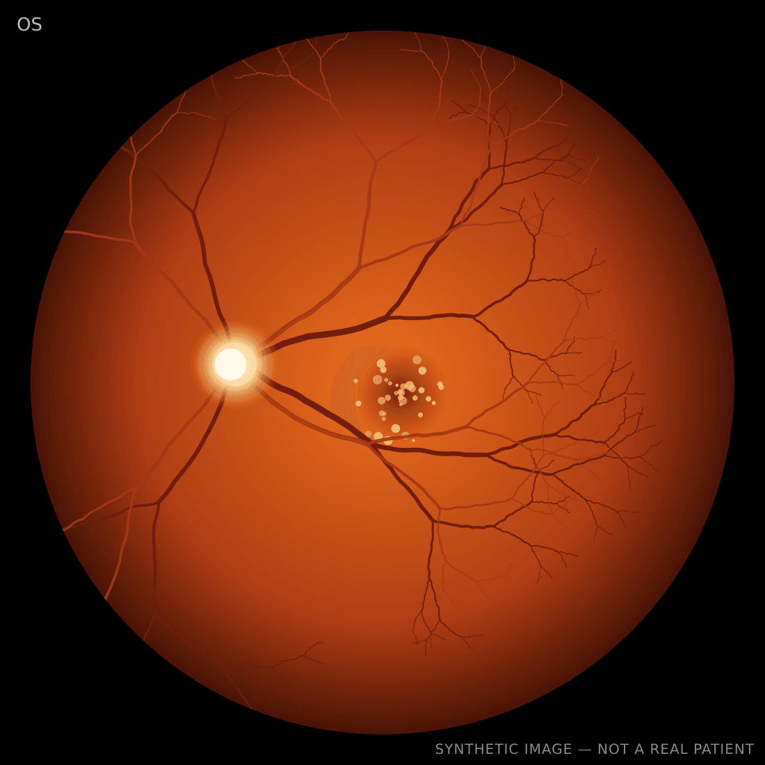
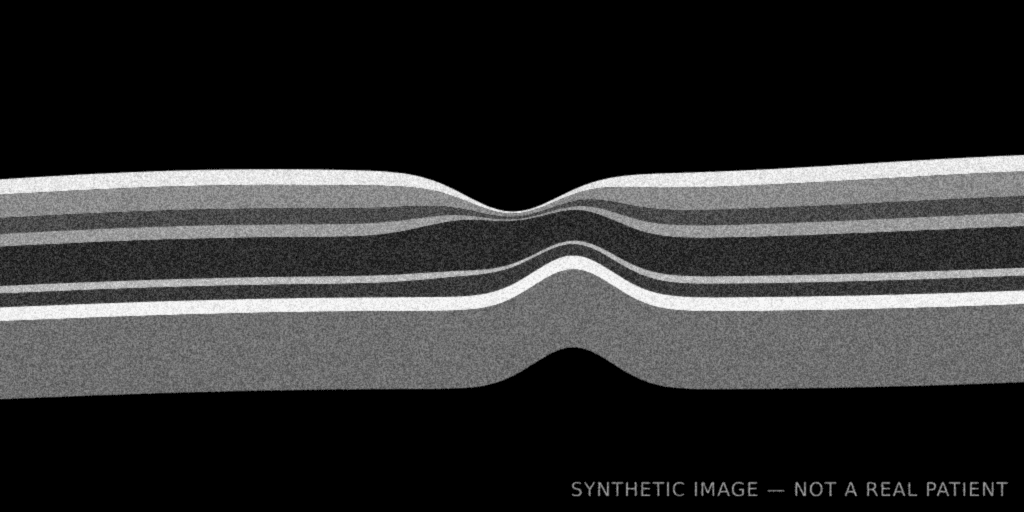
3 images
OCT captured from the workstation screen, fundus through an HDMI capture card
Drusen OS with new distortion for 3 days. Any sign of fluid, and how soon should I refer?
- OS
- Distortion
- VA 20/50
- Routine
You get back
Retina eConsult report
Final, signed
- Recommended disposition
- Retina evaluation within 1–2 weeks
- Image quality
- Diagnostic
Impression
Intermediate AMD with a small pigment epithelial elevation OS. No definite subretinal fluid on the submitted scan.
Additional evaluation
Fellow-eye OCT at the visit; return sooner if distortion worsens.
Electronically signed by the reading retina specialist. Delivered as a PDF to view, print, download or fax.
Provider to provider, by design
You remain the treating clinician
The specialist's interpretation is a consultant opinion returned to you. Clinical decisions and every conversation with the patient stay with you.
Specialists never contact patients
Readers see only the images and context you send. There are no patient accounts, no patient messaging and no prescribing through the platform.
Credentialed, state-eligible readers
Every retina specialist is verified before reading, and consults are routed only to readers licensed where your patient is seen.
How it works
Capture
Share your OCT or fundus camera window, pick an HDMI capture card, or drop files. Frames upload while you type.
Ask
One clinical question, laterality and a few symptom chips. Add more context only if you want to.
Receive
A signed PDF report you can view, print, download or have faxed to your office automatically.
For retina specialists
- See only cases you are credentialed and licensed to read, most urgent first.
- The first specialist to accept owns the case. Nobody else can open it.
- Review in a full image viewer and sign a structured report in one step.
- Message the requesting clinician when you need another scan or more context.
No installs, no proprietary hardware. If your imaging shows on screen, you can send it.
Sign in